Best Oral Radiology Treatment In Hyderabad | Alux Dental
Oral and maxillofacial radiology is the speciality of dentistry and discipline of radiology concerned with the production and interpretation of images and data produced by all modalities of radiant energy that are used for the diagnosis and management of diseases, disorders and conditions of the oral and maxillofacial region.
Radiological Imaging is an important diagnostic adjunct to the clinical assessment of the dental patient.
What are conventional X rays?
Conventional X-rays are the most common type of x-rays. There are several types of intraoral x-rays. Each one shows different aspects of the teeth.
What are Bitewing X rays?
Bite-wing x-rays show details of the upper and lower teeth in one area of the mouth. Each bite-wing shows a tooth from its crown (the exposed surface) to the level of the supporting bone.
Bite-wing x-rays detect decay between teeth and changes in the thickness of bone caused by gum disease. Bite-wing x-rays can also help determine the proper fit of a crown (a cap that completely encircles a tooth) or other restorations (eg, bridges). It can also see any wear or breakdown of dental fillings.
What are Periapical X rays?
Periapical x-rays show the whole tooth — from the crown, to beyond the root where the tooth attaches into the jaw. Each periapical x-ray shows all teeth in one portion of either the upper or lower jaw. Periapical x-rays detect any unusual changes in the root and surrounding bone structures.
What are occlusal x-rays?
Occlusal x-rays track the development and placement of an entire arch of teeth in either the upper or lower jaw.
DIGITAL X-RAYS
What is Radiovisiography?
The RadioVisioGraphy (RVG) imaging system is a form of X-ray imaging, where digital X-ray sensors are used instead of traditional photographic film. It is used to take intraoral periapical radiographs, delivering the highest image resolution.
Advantages include time efficiency through bypassing chemical processing and the ability to digitally transfer and enhance images. Also, less radiation can be used to produce an image of similar contrast to conventional radiography.
What is an OPG scan?
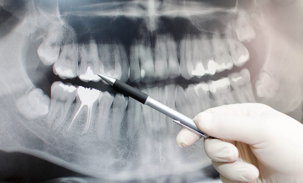
An OPG is a panoramic or wide view x-ray of the lower face, which displays all the teeth of the upper and lower jaw on a single film. It demonstrates the number, position and growth of all the teeth including those that have not yet surfaced or erupted. It is different from the small close up x-rays dentists take of the individual.
teeth. An OPG may also reveal problems with the jawbone and the joint which connects the jawbone to the head called the Temporomandibular joint or TMJ. An OPG may be requested for the planning of orthodontic treatment, for assessment of wisdom teeth or for a general overview of the teeth and the bone which supports the teeth.
How different is OPG X-ray from regular intra-oral X-ray?
Regular intra-oral x-ray provides only the selective view of particular teeth. Whereas an OPG x-ray gives the dentist a complete panoramic view of all the teeth and adjacent bones.
This aids in making well-informed decisions in a wide variety of dental cases and helps in deriving an evidence-based treatment plan.
How much time is involved in capturing an OPG X-Ray?
The process of capturing an OPG X-ray usually requires a minute or so.
What is a LATERAL CEPHALOGRAM?
A Lateral Cephalogram is a lateral or side view x-ray of the face, which demonstrates the bones and facial contours in profile on a single film. Lateral Cephalogram x-rays are usually used in the diagnosis and treatment of orthodontic problems.
DO Dental radiographs, if repeated, have harmful effects?
No, dental radiographs contribute very little radiation which is negligible when compared to the harmful dosage of other forms of radiation.
Are x- rays dangerous for pregnant women? Is it an absolute contraindication?
In pregnant patients, radiographs can be taken in emergency conditions if proper radiation safety and protective measures are taken.
3D DIMENSIONAL IMAGING:
What is a CBCT scan?
It’s basically a three-dimensional radiograph. CBCT stands for ―Cone Beam Computed Tomography. It uses a special type of technology to generate three dimensional (3-D) images of dental structures, soft tissues, nerve paths and bone in the craniofacial region in a single scan. Images obtained with Cone Beam CT allow for more precise treatment planning. There is lesser distortion as compared to 2D images.
You may be asked to get a CBCT scan done in the following cases-
- Dental implants –for the precise planning and placement of dental implants.
- For Assessment of Relative Bone Quality
- Root canal treatment- for clear-cut viewing of root canals.
- Temporomandibular Joint– for diagnosis of TMJ related problems
- Impacted teeth – for checking the position of the impacted tooth in relation to the neighbouring structures like mandibular nerve and maxillary sinus.
- Orthodontic treatment– for evaluation of the facial structures and teeth.
What are the benefits of CBCT scan over 2D radiographs?
- Benefits of CBCT scan include high definition images with exceptional details.
- Multidimensional assessment is possible as multiple images are generated in various planes.
- Helpful inaccurate diagnosis and treatment planning.
- Beneficial for Guided Surgeries.
CBCT or 3D imaging is surely going to shape the future of dental diagnosis and treatment planning. This quick and painless procedure helps inaccurate assessment of your facial skeleton and helps in the planning of reconstructive and cosmetic dentistry procedures. At ALUX DENTAL we have the latest KAVO OP 3D imaging modality; one of the best CBCT machines for our patients.


